File:Figure-8.jpg
Jump to navigation
Jump to search
Size of this preview: 800 × 247 pixels. Other resolutions: 320 × 99 pixels | 1,353 × 418 pixels.
Original file (1,353 × 418 pixels, file size: 106 KB, MIME type: image/jpeg)
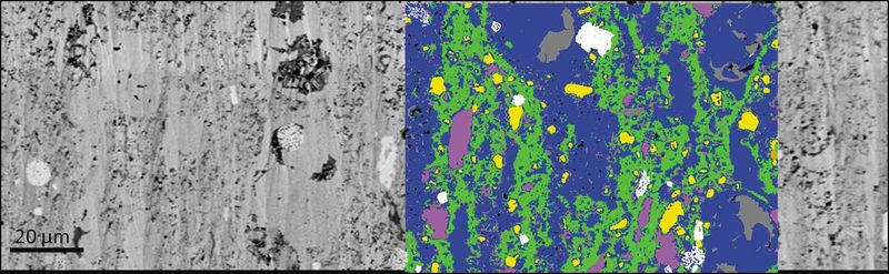
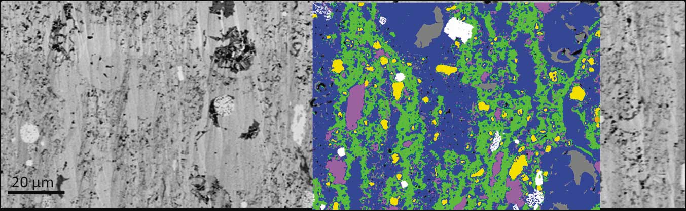
A secondary electron image of a shale sample with an EDS-derived mineral segmentation overlay. In the segmented region, blue = carbonate, green = clay minerals, yellow = quartz, pink = feldspar, white = pyrite, and gray = organic matter. From AAPG Memoir 102, Chapter 1, Huang et al., 2013, DOI: 10.1306/13391699M1023580.
File history
Click on a date/time to view the file as it appeared at that time.
| Date/Time | Thumbnail | Dimensions | User | Comment | |
|---|---|---|---|---|---|
| current | 17:34, 15 August 2014 | 1,353 × 418 (106 KB) | Molyneux (talk | contribs) | A secondary electron image of a shale sample with an EDS-derived mineral segmentation overlay. In the segmented region, blue = carbonate, green = clay minerals, yellow = quartz, pink = feldspar, white = pyrite, and gray = organic matter. From AAPG Memo... |
You cannot overwrite this file.
File usage
The following page uses this file: