Difference between revisions of "Scanning electron microscopy (SEM)"
Jump to navigation
Jump to search
Cwhitehurst (talk | contribs) |
Cwhitehurst (talk | contribs) |
||
| Line 13: | Line 13: | ||
==References== | ==References== | ||
| − | + | {{reflist}} | |
| − | |||
| − | |||
[[Category:Laboratory methods]] | [[Category:Laboratory methods]] | ||
Revision as of 20:38, 23 June 2014
Scanning electron microscopy is simply the process of using a scanning electron microscope.

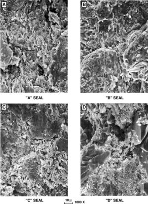
Figure 1 Images from an SEM.[1]
The scanning electron microscope became available commercially in the mid 1960s and is used by geologists to study pore geometry and diagenetic history in order to evaluate type, distribution, and flow of fluids in the lithosphere. The SEM is useful for examining the effect of fluids and chemical additives on rocks during enhanced oil recovery.[2]
Figure 1 shows images from an SEM.[1] These images are SEM photomicrographs of seal types.
This article is a stub. You can help AAPG Wiki by expanding it.
Examples of use
- Camp, W.K, E. Diaz, and B. Wawak, eds., Electron Microscopy of Shale Hydrocarbon Reservoirs: AAPG Memoir 102, 260 pp.
References
- ↑ 1.0 1.1 Snider, Robert M., John S. Sneider, George W. Bolger, and John W. Neasham, 1997, Comparison of seal capacity determinations: Conventional cores vs. cuttings, in Surdam, R. C., ed., Seals, Traps, and the Petroleum System: AAPG Memoir 67, pp. 1-12.
- ↑ Thomas, John B., and Edward D. Pittman, 1979, Applications of scanning electron microscopy to hydrocarbon exploitation: AAPG Bulletin, v. 63 No. 3, p. 539.